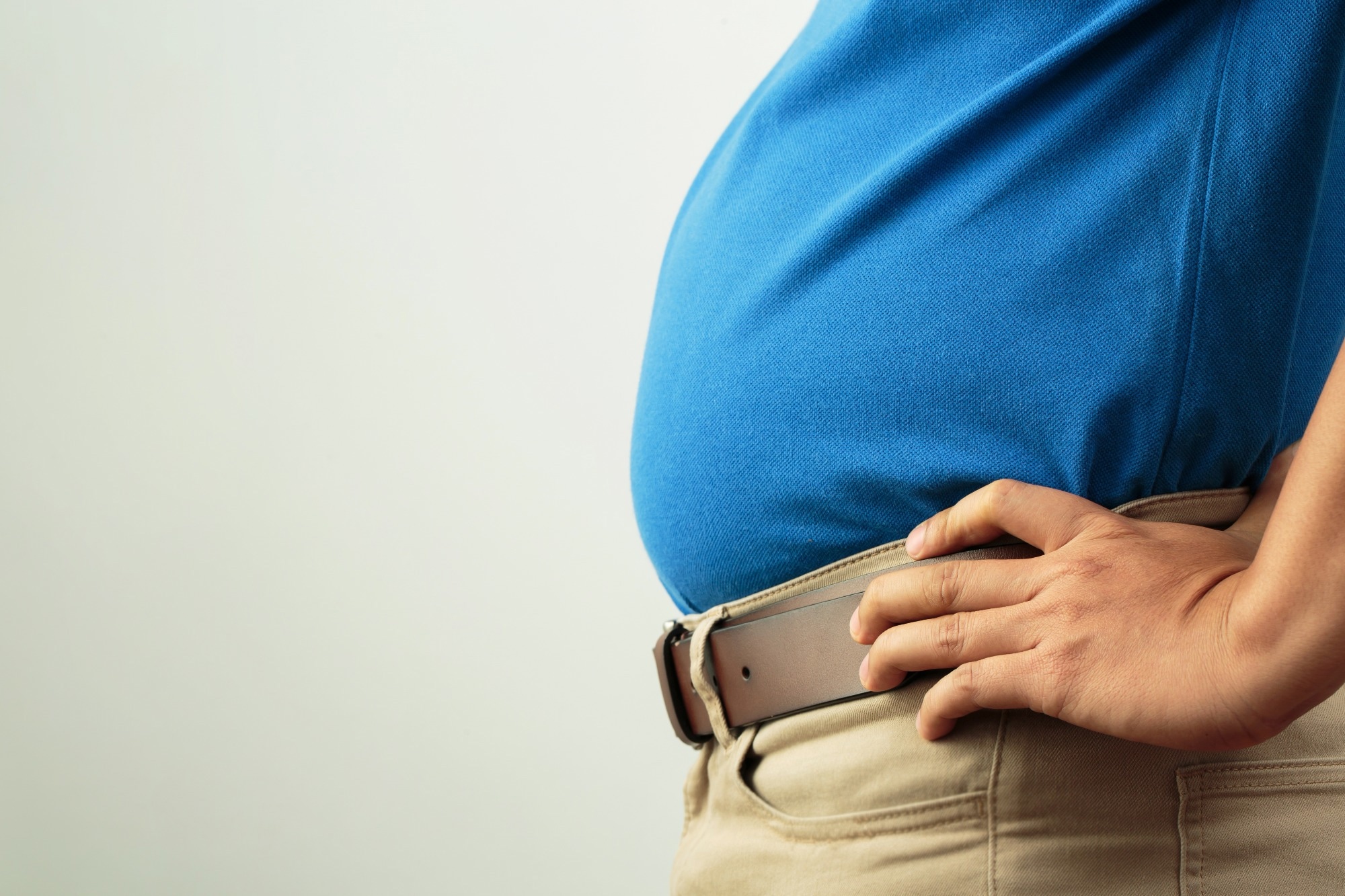
A recent study published in the journal PNAS reports that abdominal obesity is associated with excessive inflammatory responses and an increased risk of mortality in coronavirus disease 2019 (COVID-19) patients.
 Study: Apple-shaped obesity: A risky soil for cytokine-accelerated severity in COVID-19. Image Credit: fongbeerredhot / Shutterstock.com
Background
COVID-19, which is caused by infection with the severe acute respiratory syndrome coronavirus 2 (SARS-CoV-2), is a multifactorial disease. Excessive cytokine production and hyperinflammation are considered to be major hallmarks of severe COVID-19.
In addition to increasing age, various comorbidities, including hypertension, diabetes, chronic kidney disease, and obesity, increase the risk of severe COVID-19, hospitalization, and mortality. In Japan, the presence of abdominal obesity, which is defined as visceral adipose tissue-dominated obesity, is associated with poor prognosis in COVID-19 patients.
About the study
In the current study, scientists compare COVID-19 outcomes between Japanese patients with and without abdominal obesity. To this end, abdominal computed tomography (CT) imaging was used to quantify fat areas in the abdomen and subcutaneous tissues and correlate these values to COVID-19 severity and outcomes.
The researchers also investigated the characteristic features of COVID-19 in two obese mouse models. These models included ob/ob mice, which have a genetic defect in their leptin ligand, whereas db/db mice have a genetic dysfunction of the leptin receptor. As compared to db/db mice, ob/ob mice exhibit higher liver and perirenal adipose tissue weights, thus indicating that this model can be used to resemble visceral adipose tissue (VAT)-dominant obesity.
Important observations
Increased VAT accumulation was found to be an independent risk factor for COVID-19 mortality, even when other factors such as old age, a history of infarction, and chronic kidney disease (CKD) were also present. Patients with VAT-dominated obesity, which can otherwise be described as abdominal obesity, were also significantly more susceptible to severe COVID-19 and mortality as compared to those with high body mass index (BMI) or subcutaneous adipose tissue (SAT)-dominated obesity.
Increased adipose tissue was also weakly associated with C-related protein (CRP), D-dimer, and ferritin levels at both early and peak stages of the disease. The higher concentration of macrophages and adipocytes in VAT also led to a hyperinflammatory response and, as a result, worse clinical outcomes, particularly among young patients.
The in vivo experiments revealed that exposure to SARS-CoV-2 increases the risk of mortality in ob/ob mice, whereas all db/db and control mice survived. Notably, ob/ob mice also exhibited relatively higher numbers of SARS-CoV-2-positive cells in the alveoli as compared to db/db and control mice; however, SARS-CoV-2-positive cells within the bronchi were comparable between each group.
A higher susceptibility to SARS-CoV-2-related mortality was also observed in high-fat diet (HFD)-fed mice as compared to that in normal fat diet (NFD)-fed mice. HFD-fed mice also exhibited similar immunopathological findings as those observed in ob/ob mice with abdominal obesity.
A high level of inflammation and acute lung injury was observed in HFD-fed mice and ob/ob mice. In contrast, mice with SAT-dominated obesity exhibited localized inflammation and attenuated lung injury upon SARS-CoV-2 exposure, thereby indicating favorable COVID-19 outcomes.
An increased proportion of SARS-CoV-2 antigen-positive lung macrophages was observed in mice with abdominal obesity. Electron microscopic analysis identified disrupted viral particles in these macrophages. Further analysis revealed an upregulated type I/II interferon response and interleukin-6 (IL-6) level in the lungs of abdominal obesity mice.
Ribonucleic acid (RNA) sequencing analysis identified an enrichment of inflammation-related pathways in the lungs of abdominal obesity mice. These included cytokine-cytokine receptor interaction, tumor necrosis factor (TNF) signaling, chemokine signaling, and nucleotide-binding, and oligomerization domain (NOD)-like receptor pathways.
These observations collectively indicate that viral antigen-carrying lung macrophages circulate systemically and induce inflammatory responses in mice with abdominal obesity. Increased inflammation characterized by a cytokine storm might be responsible for higher mortality in SARS-CoV-2-infected mice with abdominal obesity.
Treatment of these mice with IL-6 receptor-targeting antibodies revealed a significant reduction in mortality rates, which further confirms the association between increased cytokine production and higher mortality risk.
Adipose tissue modification
The current study explored the extent to which obesity influences COVID-19 outcomes in mice treated with leptin before viral exposure. To this end, leptin-mediated reduction in body weight led to improved survival rates after infection in mice with abdominal obesity.
Prophylactic leptin administration also led to a reduction in inflammatory lung macrophages, viral RNA load, IL-6 production, interferon response, and adipose tissue weight in these mice. An attenuation in inflammation-related pathways and improvement in lysosomal function of alveolar macrophages were observed in mice that received preventive leptin.
Thus, excessive accumulation of adipose tissue in the abdomen is associated with delayed viral clearance, activation of inflammatory macrophages and cytokine production, and increased mortality in SARS-CoV-2-infected mice.
Study significance
The study findings indicate that abdominal obesity is a major risk factor for COVID-19, as it can significantly increase the risk of cytokine storm and mortality in patients infected with SARS-CoV-2. Based on these findings, COVID-19 patients with abdominal obesity might benefit from cytokine-suppressing therapies during the disease course.
Journal reference:
- Hosoya, T., Oba, S., Komiya, Y., et al. (2023). Apple-shaped obesity: A risky soil for cytokine-accelerated severity in COVID-19. PNAS. doi:10.1073/pnas.2300155120
