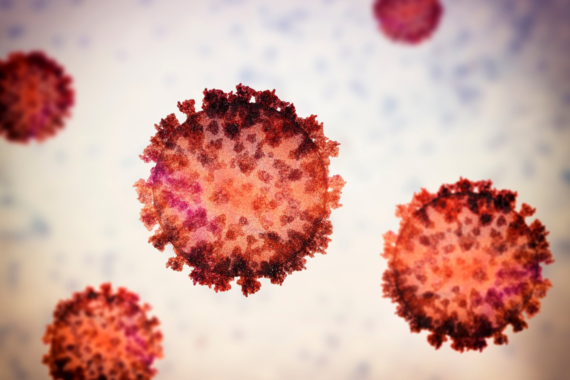
A latest examine revealed within the Science Advances Journal demonstrated that extreme acute respiratory syndrome coronavirus 2 (SARS-CoV-2) an infection and viral fusogens trigger glial and neuronal fusion.
 Research: SARS-CoV-2 an infection and viral fusogens trigger neuronal and glial fusion that compromises neuronal exercise. Picture Credit score: KaterynaKon/Shutterstock.com
Background
Fungi, micro organism, viruses, and parasites could cause nervous system infections. Viruses corresponding to Zika virus, reovirus, SARS-CoV-2, and herpes simplex virus can infect neurons.
Viral mind infections can result in fever, lack of scent or style, epileptic seizures, confusion, complications, and in extreme circumstances, meningitis, encephalitis, paralysis, and demise could happen.
In non-neuronal tissues, reoviruses and enveloped viruses use fusogens to fuse with host mobile membranes, enter cells, and hijack mobile equipment to synthesize viral parts.
The newly generated fusogens redecorate the cell membrane, imparting the power to fuse with adjoining cells, leading to multinucleated syncytia. Whether or not viral an infection and fusogens trigger, neuronal fusion and syncytia formation stay unknown.
The examine and findings
Within the current examine, researchers investigated whether or not SARS-CoV-2 an infection and viral fusogens trigger neuronal cell fusion. First, a inhabitants of mouse mind cells was transfected with plasmids encoding human angiotensin-converting enzyme 2 (hACE2) and inexperienced fluorescent protein (GFP).
A second inhabitants was transfected with plasmids encoding hACE2 and mCherry. These populations had been co-plated and maintained in vitro for 5 days.
Cultures had been contaminated with 20, 2 x 10³, or 2 x 10⁶ plaque-forming items of ancestral SARS-CoV-2. The cultures had been examined 72 hours post-infection by confocal microscopy. Fused neurons had been noticed, and antibody staining confirmed that the cells had been spike-positive. Additional, the workforce noticed further fusion phenotypes, corresponding to glia-glia and glia-neuron fusions.
Cell harm was noticed at larger SARS-CoV-2 titers solely. Subsequent, mind organoids derived from human embryonic stem cells (hESCs) had been contaminated with SARS-CoV-2. Likewise, these contaminated organoids confirmed neuronal syncytia.
Moreover, to judge the consequences of fusogens alone, the workforce used a SARS-CoV-2 spike and the p15 fusion protein of baboon orthoreovirus.
One inhabitants of mice neurons was transfected with p15- and GFP-expressing plasmids. One other inhabitants was transfected with a mCherry plasmid and an empty vector. The populations had been co-plated and maintained in tradition. p15 expression was ample to trigger neuronal fusion, which was not noticed with out p15.
Utilizing an inactive p15 fully abolished fusion. Additional, the researchers repeated these experiments for the SARS-CoV-2 spike protein. One neuronal inhabitants was transfected with spike- and GFP-expressing plasmids; one other was transfected with mCherry- and hACE2-expressing plasmids.
The populations had been co-plated and maintained in tradition for 72 hours. Fusion of cells expressing spike with hACE2-expressing cells was noticed. Each hACE2 and spike had been obligatory for fusion. Utilizing inactive spike variations didn’t induce fusion. Glia-glia and glia-neuron fusion phenotypes had been noticed with each p15 and spike fusogens.
Subsequent, the workforce investigated whether or not fusogens might induce fusion in vivo. To this finish, vectors expressing GFP alone or GFP and p15 had been injected into the cortex and hippocampus of 11-week-old mice, and brains had been eliminated after seven and 14 days. Neuronal fusion was detected within the cortex and the hippocampus.
They subsequent examined in human-derived neurons with SARS-CoV-2 spike or p15. hESC-derived cortical neurons and neuronal progenitor cells had been transfected with GFP and spike, inactive spike, or p15. Three days later, clusters of interconnected GFP-positive neuronal cells had been evident with p15, much like the syncytia noticed in murine neurons.
Clusters had been additionally fashioned when the SARS-CoV-2 spike was expressed, implying that fusion might happen by using endogenous hACE2 receptors. No fusion was detected with the inactive spike or with out fusogens. Subsequent, the researchers explored whether or not fusion was potential on the stage of neurites, away from the cell our bodies.
SARS-CoV-2 an infection induced fusion between somas and between neurites distant from somas. Fusion resulted in bridges of various lengths exceeding lots of of micrometers. The workforce noticed the trade of mitochondria and a fluorescent protein (mCardinal) by way of the fusion bridges.
Additional, neurons transfected with GFP and p15 had been noticed for syncytia formation over seven days. Syncytia elevated over time within the presence of p15, with the progressive incorporation of latest cells. Lastly, the researchers examined whether or not fusion impacted neuronal exercise.
To this finish, differentiated neurons had been fused utilizing p15, and fusion was visualized utilizing a fluorescent indicator delicate to calcium ions. Neurons expressing mCherry and empty vector had spontaneous exercise. Most fused neurons (90%) confirmed synchronized neuronal exercise, whereas the remaining confirmed full lack of neuronal exercise.
Cells with out exercise had been people who fused tightly on the stage of somas. All neurons fused with glia had misplaced exercise. The frequency of the neuronal exercise was unaltered, whatever the synchronicity of fused neurons. The workforce additionally noticed elevated intracellular calcium ion ranges throughout the bridges.
Conclusions
The findings demonstrated that virus-infected neurons or these expressing viral fusogens might fuse with adjoining neurons and glia, resulting in adjustments in neuronal communication and compromising neuronal exercise. Neuronal fusion by SARS-CoV-2 is dependent upon hACE2 expression and, presumably, different accent elements.
Fusion additionally resulted within the trade of huge molecules and organelles. Furthermore, fused neurons had been viable however with altered perform and circuitry. Most current SARS-CoV-2 vaccines, together with BNT162b2, ChAdOx1, mRNA-1273, and Ad26.COV2.S are primarily based on spike expression in host cells to elicit the immune response.
These vaccines encode the full-length spike with two mutations stabilizing the prefusion conformation and inactivating their fusogenicity. The researchers used the identical model of the inactive spike within the present investigation, which did not set off fusion.
The findings recommend that contemplating the fusogenic potential will probably be essential in designing future SARS-CoV-2 vaccines.
